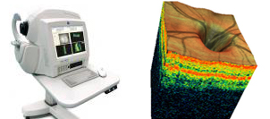
Glaucoma Diagnosis Advances With Ocular Coherence Tomography (OCT)
The Ocular Coherence Tomographer (OCT) provides high resolution cross-sectional imaging of ocular tissues. It can measure retinal nerve fibre layer tissue as will as macula thickness and can be used to study glaucoma as well as macular conditions such as macular degeneration and edema.

The technique is analagous to ultrasound except is uses light (843nm) instead of sound waves to allow exceptionally high resolution. Reflected light from nerve or retinal tissue and a reference mirror interact to produce an interference pattern which is detected and processed into a signal. Strong reflections occur at the interface between materials of different refractive indices and from tissues with high scattering coefficients. Distances between ocular tissues can be measured from the time taken for the light to reflect from the various structures within the eye. A tomographical image is generated by simultaneously performing and displaying 100 longitudinal adjacent scans. Image quality can be affected by such things as cataracts but are still obtainable in most situations.
Back